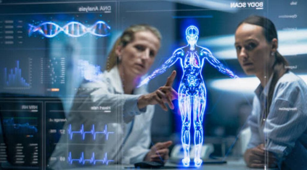
Artificial intelligence that can “read” electronic radiology reports and flag patients with broken bones who are at risk of osteoporosis outperformed the traditional manual method of health care professionals reading X-ray reports, a new study finds.
The results were presented during a virtual news conference hosted by the Endocrine Society.
The new search tool, called X-Ray Artificial Intelligence Tool (XRAIT), detected an almost fivefold higher number of major fractures, or bone breaks, in X-ray and computed tomography (CT) reports compared with manual methods, researchers from Australia reported.
“With XRAIT, limited health care resources can be optimized to manage the patients identified as at risk rather than used on the identification process itself,” says study co-investigator Jacqueline Center, MBBS, PhD, FRACP, the head of the Clinical Studies and Epidemiology Lab at Garvan Institute of Medical Research in Sydney, Australia. “By improving identification of patients needing osteoporosis treatment or prevention, XRAIT may help reduce the risk of a second fracture and the overall burden of illness and death from osteoporosis.”
About 44 million Americans—mostly women—are at risk of the bone-weakening disease osteoporosis and have an increased risk of fractures because of low bone mass, according to the Hormone Health Network. Only two in 10 older women in the United States who sustain a fracture receive testing or treatment for osteoporosis, the National Osteoporosis Foundation reports.
Although many hospitals have implemented fracture liaison services to identify patients whose fractures could be due to osteoporosis, Center says that manually reading the radiology records of referred patients misses some people at risk of osteoporosis or detects them too slowly. XRAIT speeds the process using natural language processing software to “understand” human language.
In this study, XRAIT searched 5,089 digital radiology reports from patients over 50 years of age who went to a hospital’s emergency department and had bone imaging over three months. The researchers—led by senior author Christopher White, MBBS, PhD, FRACP, of Prince of Wales Hospital in Randwick, Australia—compared XRAIT’s results with manual review of the records of the 224 patients referred to the hospital’s fracture liaison service in the same period. XRAIT was able to detect 349 people with fractures likely due to low bone mass compared with 98 people identified by the manual method, an over three-fold higher detection rate.
Next, the researchers tested XRAIT on the digitized radiology reports of another population of Australian adults over age 60 from the Dubbo Osteoporosis Epidemiology Study. From 327 reports of confirmed known fractures and nonfractures, XRAIT accurately identified fractures nearly seven of 10 times and correctly screened out patients without fractures more than nine of 10 times, Center says. She says this finding suggests that other hospitals can easily use XRAIT. South Eastern Sydney Local Health District developed XRAIT, using software licensed to AbbottDiagnostics. Australia’s Prince of Wales Hospital Foundation gave seed funding for XRAIT.
As of now, about 80% of patients with osteoporosis leave the hospital untreated, and preventable fractures burden health systems that repair them, White says. “The process of treating osteoporosis is fractured,” he says.